Epigenetics: Linking environmental factors and gene regulation
A technique called mSTARR-seq sheds light on how DNA methylation may shape responses to external stimuli by altering the activity of sequences that control gene expression.
- Department of Genes and Environment, Max Planck Institute of Psychiatry, Germany
Gene regulation is a complex process that allows cells to control when and how they express their genes. Genetic variants can influence these mechanisms, often causing issues that lead to disease (Albert and Kruglyak, 2015). But could environmental signals also play a similar role?
This question has fuelled much interest into epigenetics, a field that focuses on a range of molecular mechanisms which modify DNA while leaving the underlying genetic sequence intact. The best-known example, DNA methylation, is crucial for fundamental biological processes such as cell fate decision. This process relies on methyl groups being added to certain genetic sites. These ‘marks’ can persist through division and therefore be transmitted across cell generations and importantly, environmental factors can influence them (Moore et al., 2013). As such, DNA methylation represents a potential mechanism by which the environment can shape gene expression and subsequent health outcomes. Indeed, early changes in DNA methylation have been proposed to dictate how an organism responds to stressors later in life, enabling adverse childhood experiences to become biologically ‘embedded’ by leaving genetic traces with life-long consequences (Aristizabal et al., 2020).
Many human studies have tried to identify the DNA methylation signatures associated with early exposure to adverse events in peripheral tissues such as blood or buccal cells (Cecil et al., 2020). As the functional impact of these changes has rarely been explored, however, it remains unclear whether they can impact future responses to stimuli. Overall, the extent to which DNA methylation affects gene expression is still poorly understood. Some studies report that methylation marks disrupt transcription factor binding and other gene expression mechanisms, but other evidence indicates they may have no effect on the activity of most regulatory elements (Kreibich et al., 2023). These conflicting results highlight the need to closely examine which type of impact methylation may have on various regulatory sites. Now, in eLife, Rachel Johnston, Jenny Tung and colleagues at Duke University and other institutes in Canada, Germany and the United States report having used a technique called mSTARR-seq to investigate the effect of DNA methylation on the activity of millions of sequences across the human genome, particularly in response to environmental factors (Johnston et al., 2024).
The team had previously developed this approach to assess the functional effects of DNA methylation on a large number of sequences ‘in one go’, but they had not examined then the impact of external stimuli. In their latest study, they aimed to address this gap by first using mSTARR-seq to assess the activity on a genome-wide level, including the most studied methylation sites in both their methylated and unmethylated states. This revealed that differences in methylation status impacted the activity of almost half of known regulatory regions, suggesting a genome-wide role of DNA methylation in gene regulation.
Next, Johnston et al. assessed whether pre-existing DNA methylation status influenced how cells respond to stimuli. To do so, they applied mSTARR-seq to immune cells exposed to molecules known to modulate inflammation and mediate stress responses. By modelling immune system activation and stress responses, the team highlighted thousands of regulatory regions that respond differentially to the compounds depending on their initial DNA methylation status. Further experiments using macrophages from human donors confirmed that differences in pre-existing methylation patterns predicted responses to viral infection (Figure 1).
Figure 1
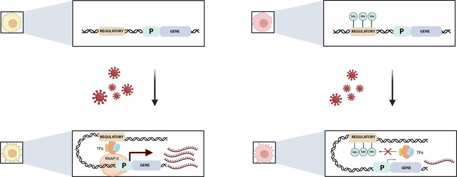
Pre-existing DNA methylation influences how macrophages respond to infection.
Viral exposure activates macrophages and leads to changes in their gene expression. Johnston et al. show that the methylation status of certain sequences prior to infection can alter this response. For example (top left), an unmethylated regulatory element (brown) controlling a gene (blue) and its associated promotor (‘P’; green) is free to recruit transcription factors (TFs) after viral exposure (bottom left). This helps a complex known as RNAPII to bind to the promotor, allowing transcription to start (maroon arrow) and large numbers of mRNA transcripts to be produced (maroon). If the regulatory element is methylated, however (top right), the methylated groups (green circles labelled Me) interfere with the recruitment of the transcription factors (red cross, bottom right) and therefore with the binding of RNAP II. This impairs transcription and the associated production of mRNA transcripts.
© 2024, BioRender Inc. Figure 1 was created using BioRender, and is published under a CC B-NC-ND license. Further reproductions must adhere to the terms of this license.
Finally, the team explored whether methylation changes linked to early life stressors could influence gene regulation. To do so, they used data from 27 studies and compiled a list of genomic regions whose methylation levels are associated with adverse childhood events. Except for one study, these sequences were not more likely to be gene regulatory regions compared to chance; they were also not enriched in sites that the mSTARR-seq analyses highlighted as displaying DNA-methylation-dependent activity. This led Johnston et al. to suggest that DNA methylation marks linked to childhood adversity in peripheral tissues might serve as indicators of exposure to early stressors, rather than cause gene expression changes with long-lasting effects.
It is important to note, however, that these findings were established by comparing data from different cell types – the mSTARR-seq analyses are based on immune cell lines, while the studies examining DNA methylation and early stressors used blood, saliva or buccal samples. Yet the physical and psychological outcomes associated with early adverse experiences involve many tissues, which are known to present different methylation patterns (Eriksson et al., 2014; Rahman and McGowan, 2022). It is therefore possible that these marks have a more causal role in other cell types. Further studies investigating how DNA methylation impacts gene expression across tissues would help to clarify the connection between stress in early life, epigenetic changes, and later health outcomes.
Our interaction with our environment is not a passive process – not even at the level of gene expression. The work by Johnston et al. helps to dissect the complexity of this relationship, highlighting how DNA methylation modulates our response to external stimuli. Their study also suggests that we should carefully interpret the role this epigenetic process plays in the long-term impact of early stressors. Numerous questions remain, including about how these findings could be translated into new targets for preventing and treating disease.
References
-
The role of regulatory variation in complex traits and diseaseNature Reviews Genetics 16:197–212.https://doi.org/10.1038/nrg3891
-
Childhood maltreatment and DNA methylation: a systematic reviewNeuroscience and Biobehavioral Reviews 112:392–409.https://doi.org/10.1016/j.neubiorev.2020.02.019
-
Early life stress and later health outcomes—findings from the Helsinki Birth Cohort StudyAmerican Journal of Human Biology 26:111–116.https://doi.org/10.1002/ajhb.22502
-
DNA methylation and its basic functionNeuropsychopharmacology 38:23–38.https://doi.org/10.1038/npp.2012.112
-
Cell-type-specific epigenetic effects of early life stress on the brainTranslational Psychiatry 12:326.https://doi.org/10.1038/s41398-022-02076-9
Article and author information
Author details
Elisabeth B Binder

Publication history
- Version of Record published: March 18, 2024 (version 1)
Copyright
© 2024, Penner-Goeke and Binder
This article is distributed under the terms of the Creative Commons Attribution License, which permits unrestricted use and redistribution provided that the original author and source are credited.
Metrics
-
- 1,247
- views
-
- 130
- downloads
-
- 0
- citations
Views, downloads and citations are aggregated across all versions of this paper published by eLife.
Download links
A two-part list of links to download the article, or parts of the article, in various formats.
Downloads (link to download the article as PDF)
Open citations (links to open the citations from this article in various online reference manager services)
Cite this article (links to download the citations from this article in formats compatible with various reference manager tools)
Epigenetics: Linking environmental factors and gene regulation
eLife 13:e96710.
https://doi.org/10.7554/eLife.96710
Further reading
-
- Chromosomes and Gene Expression
- Immunology and Inflammation
Ikaros is a transcriptional factor required for conventional T cell development, differentiation, and anergy. While the related factors Helios and Eos have defined roles in regulatory T cells (Treg), a role for Ikaros has not been established. To determine the function of Ikaros in the Treg lineage, we generated mice with Treg-specific deletion of the Ikaros gene (Ikzf1). We find that Ikaros cooperates with Foxp3 to establish a major portion of the Treg epigenome and transcriptome. Ikaros-deficient Treg exhibit Th1-like gene expression with abnormal production of IL-2, IFNg, TNFa, and factors involved in Wnt and Notch signaling. While Ikzf1-Treg-cko mice do not develop spontaneous autoimmunity, Ikaros-deficient Treg are unable to control conventional T cell-mediated immune pathology in response to TCR and inflammatory stimuli in models of IBD and organ transplantation. These studies establish Ikaros as a core factor required in Treg for tolerance and the control of inflammatory immune responses.
-
- Cell Biology
- Chromosomes and Gene Expression
Heat stress is a major threat to global crop production, and understanding its impact on plant fertility is crucial for developing climate-resilient crops. Despite the known negative effects of heat stress on plant reproduction, the underlying molecular mechanisms remain poorly understood. Here, we investigated the impact of elevated temperature on centromere structure and chromosome segregation during meiosis in Arabidopsis thaliana. Consistent with previous studies, heat stress leads to a decline in fertility and micronuclei formation in pollen mother cells. Our results reveal that elevated temperature causes a decrease in the amount of centromeric histone and the kinetochore protein BMF1 at meiotic centromeres with increasing temperature. Furthermore, we show that heat stress increases the duration of meiotic divisions and prolongs the activity of the spindle assembly checkpoint during meiosis I, indicating an impaired efficiency of the kinetochore attachments to spindle microtubules. Our analysis of mutants with reduced levels of centromeric histone suggests that weakened centromeres sensitize plants to elevated temperature, resulting in meiotic defects and reduced fertility even at moderate temperatures. These results indicate that the structure and functionality of meiotic centromeres in Arabidopsis are highly sensitive to heat stress, and suggest that centromeres and kinetochores may represent a critical bottleneck in plant adaptation to increasing temperatures.


